How to Reliably Prove the Efficacy of a Toxin Binder
Proving feed additive efficacy in binding mycotoxins requires more than a single test. This article outlines how a combination of in vitro binding assays and a controlled broiler trial delivers the scientific evidence needed to validate a toxin binder with confidence.
Mycotoxins pose a serious and widespread threat to animal nutrition, causing substantial economic damage across the global livestock industry. To counter their harmful effects, the neutralization of mycotoxins has become a standard tool in modern feed management, yet demonstrating the true efficacy of these products remains a significant challenge.
A reliable starting point is the assessment of adsorption capacity under standardized conditions. These in vitro studies offer an objective and reproducible first screening, allowing for a meaningful comparison of mycotoxin binder products. But laboratory results alone are not enough: what ultimately matters is measurable performance in living animals.
This article presents a rigorous two-step approach to evaluating toxin binder efficacy, beginning with in vitro testing and culminating in a controlled scientific broiler trial.
In vitro study
Specific in vitro studies were developed to replicate the physiological conditions found along an animal's digestive tract. In the first phase, the adsorption of a defined mycotoxin concentration by a toxin binder product (B.I.O.Tox®, Biochem) was measured after three hours at an acidic pH of 3, mimicking the harsh environment of the stomach. The process then continued into a simulated intestinal phase, where desorption was recorded after another three hours at a neutral pH of 6.5. The final binding efficiency is derived by subtracting the desorption value from the adsorption value, yielding a clear and comparable measure of the product's true adsorption capacity.
The mean efficiency rates (n=3) are shown in Figure 1. The product showed high binding efficiency across all tested mycotoxins at the applied concentrations.
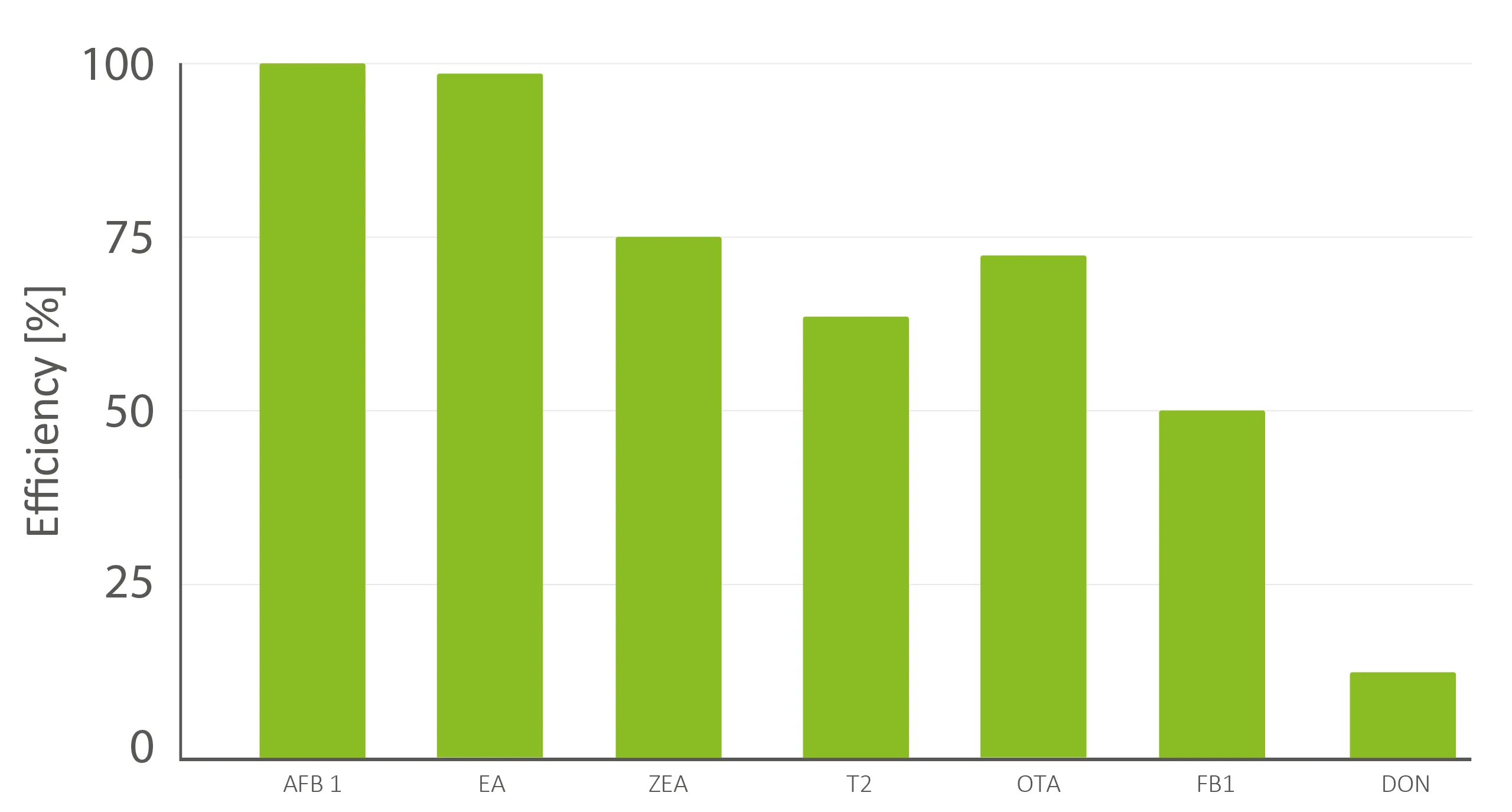 Figure 1: In vitro binding efficiency (adsorption-desorption) of AFB1 (20 ppb), EA (300 ppb), ZEA (450 ppb), T-2 (150 ppb), OTA (30 ppb), FB1 (1,000 ppb), DON (450 ppb) and an inclusion level of 1.0 kg/ton B.I.O.Tox®.
These promising in vitro results, however, must ultimately be confirmed under the complex physiological conditions of a living animal.
In vivo broiler trial
Under real-world conditions, feed ingredients are rarely contaminated with just one mycotoxin. Multiple moulds often co-occur, producing an unpredictable cocktail of toxins whose combined effects can be additive or even synergistic. The following scientific broiler trial, conducted at an independent scientific trial facility, simulates exactly this scenario: a high-level multi-mycotoxin challenge, designed to evaluate the physiological damage caused by the mycotoxins as assessed through histopathological, serological, and zootechnical parameters. Additionally, the trial investigated whether the toxin binder (BT) can effectively mitigate this systemic damage.
The trial was performed on 1,350 day-old Cobb 500 male broiler chicks, randomly allocated to five feeding groups (Table 1).
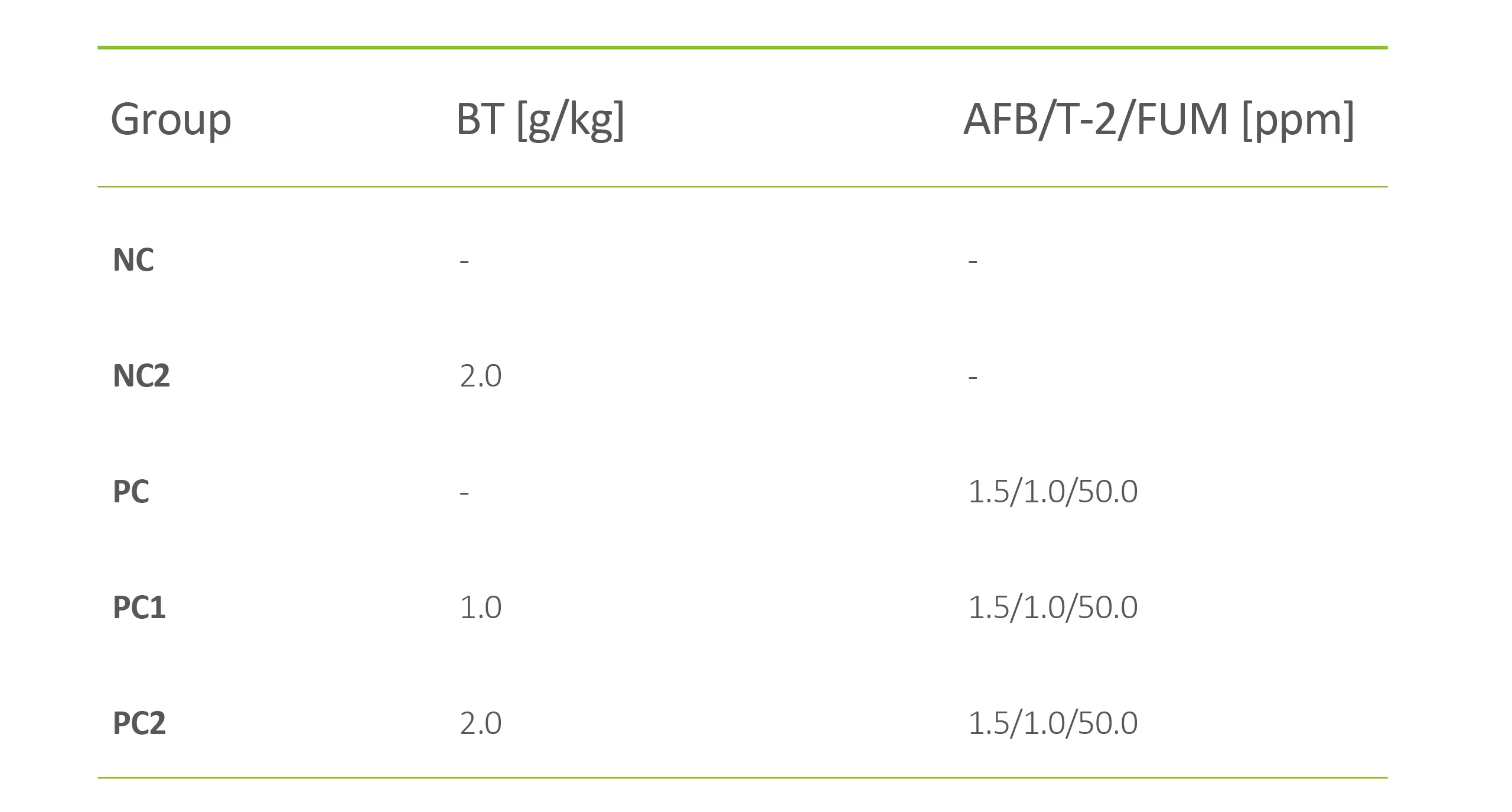 Table 1: Experimental feeding groups with the concentrations of toxin binder (BT) and mycotoxins (AFB, T-2, FUM).
All feeding groups received isonutritive diets formulated in line with the Cobb Broiler Management Guide, based on NIRS evaluation and composed of maize, soybean meal, and vitamin/mineral premix. To ensure experimental integrity, the complete trial diets were screened for mycotoxin contamination, but none was detected.
Each group comprised 270 birds consisting of nine repetitions with 30 chicks per box kept in a commercial house on rice husk litter. Trial duration was 35 days, and the following parameters were collected:
Zootechnical performance is represented as average per group by feed intake, body weight, body weight gains, and feed conversion ratio (FCR).
Histomorphology of the jejunum is shown by the villus height/crypt depth ratio (VH/CD ratio)
Relative liver weights (RLW) calculated by liver weight/carcass weight x 100, given as mean per group
Histopathology of the liver shown by periportal heterophilic inflammatory infiltration with the following scoring: absent (score 0), mild (score 1), moderate (score 2), marked (score 3)
Histopathology of the bursa of Fabricius, shown by lymphoid depletion and abscesses, applying the same scoring as for the liver
oxidative stress measured by thiobarbituric acid reactive substances (TBARS), nmol MDA (malondialdehyde)/mg protein calculated as average per group
liver function derived from total plasma protein (TP), g/dl, calculated as the average per group
The following results were observed:
Zootechnical parameters:
Feed intake, body weights and body weight gains were significantly lower in PC compared to NC and NC2. In PC1 and PC2, all three parameters were significantly lower than in NC and NC2, though being numerically or significantly higher than in PC in a dose-dependent manner (Figure 2).
No significant differences in FCR were observed between groups.
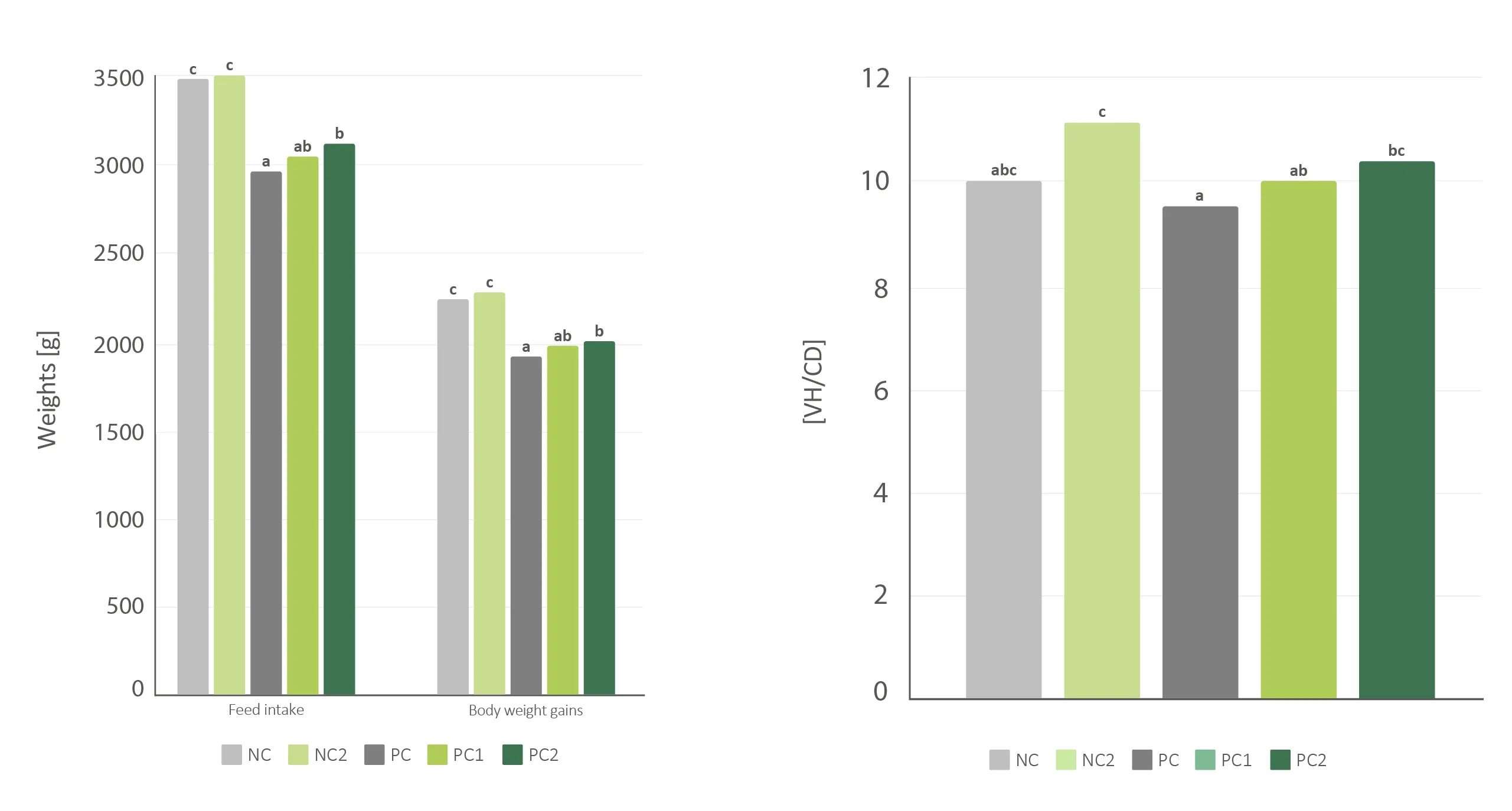 Figure 2: Performance (left) and villus height to crypt depth ratio (VH/CD) (right); a-c Values labelled with a different superscript within a row differ significantly (p<0.05).
Histopathological parameters:
The VH/CD ratio was numerically lower in PC compared to NC and significantly lower compared to NC2, indicating the adverse effect of the mycotoxins on gut epithelium. This reduction was successfully prevented in PC2 (Figure 2).
Relative liver weights were significantly higher in PC compared to NC and NC2, indicating hepatotoxic effects. For PC1 and PC2, the weights were numerically lower than those of PC.
Heterophilic infiltration in the liver was higher in PC with a median score of two and a median score of one in PC1 and PC2 compared to zero in NC and NC2. Distribution of the single scores showed milder lesions in PC1 and PC2 than in PC, showing on the one hand the hepatic damage caused by the applied mycotoxins and on the other hand the protective effect of the toxin binder.
In the bursa of Fabricius, lymphoid depletion was clearly higher in PC with a median score of one compared to zero in NC, NC2, PC1 and PC2. Regarding abscesses, all five groups kept a median score of zero, although PC revealed the highest grades detected, showing the mycotoxin caused immunosuppression (Figure 3).
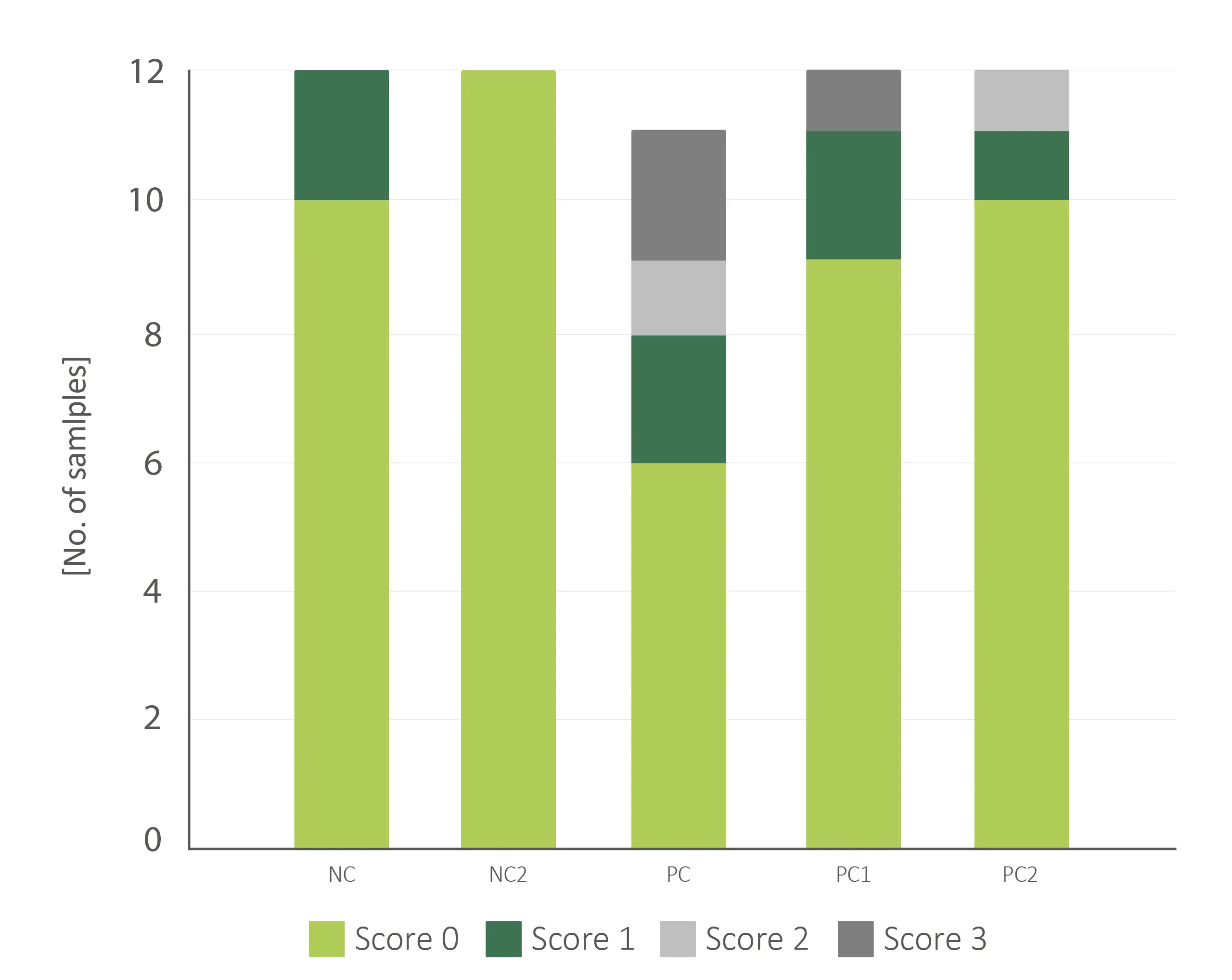 Figure 3: Abscesses in the bursa of Fabricius; one sample was discarded in PC.
Serological parameters:
TBARS were significantly higher in PC compared to NC and NC2, indicating a high oxidative stress level due to the mycotoxins. This significant increase was fully prevented in PC1 and PC2 (Figure 4).
TP was numerically lower in PC compared to NC and NC2 and was prevented in PC1 and PC2 (Figure 4), indicating an impaired protein synthesis in the liver.
 Figure 4: TBARS/TP; a-b Values labelled with a different superscript within a row differ significantly (p<0.05).
Conclusion
The mycotoxins applied in this trial caused severe and measurable damage across all evaluated parameters, including histopathological lesions, impaired serological markers, and significantly reduced zootechnical performance. The toxin binder demonstrated strong and consistent efficacy, not only in capturing mycotoxins under controlled in vitro conditions, but also in a comprehensive in vivo trial. It effectively mitigated the adverse effects of multi-mycotoxin contamination on gut morphology, liver and bursa function, and oxidative stress levels in broilers. Taken together, the findings suggest that the product successfully neutralized the applied mycotoxins before measurable damage could occur in the investigated tissues and organs, a powerful validation of the two-step evaluation approach.


